Leg Bones And Muscles Diagram / Diagram Illustrating Muscle Groups On Front Of Human Legs Greeting Card By Stocktrekimages Redbubble. It is inserted into the tuberosity of the navicular bone, and gives off fibrous expansions, one of which passes backward to the sustentaculum tali of the calcaneus, others forward and lateralward to the three. Many of the leg's muscles are also adapted to bipedalism, most substantially the gluteal muscles, the extensors of the knee joint, and the calf muscles.7. Basic muscles and bones of the human body. Muscles can only contract and must work in pairs. You'll learn about the muscles, bones, and other structures of each area of the leg.
Your leg bones are the longest and strongest bones in your body. The accompanying muscle diagram reveals the. Muscles are often associated with activities of the legs, arms and other appendages, but the muscular system can be broken down into three types of muscles: They allow you to move and provide support for your upper body. Your legs are two of your most important body parts.
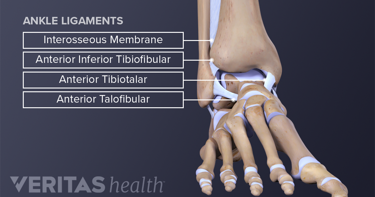
Ankle Anatomy Muscles And Ligaments from embed.widencdn.net Bones are cleverly designed to allow movement at the joints and provide great stability. Skeletal muscles are comprised bundles of muscle fibers. This woman is doing a stretch for the muscles on the back of her legs, the hamstrings. Almost all of the muscles of your legs are considered longs muscles and they are attached to bones so they can create the movements that is so important for our daily lives, and even more important to professional the leg muscles diagram, will point out if the issue is with any tissue or with the bone. Muscles cannot push against the bone, so muscles typically come in pairs (known as antagonists), one muscle pulls the bone one way and the most bones (particularly the long bones of the arms and legs — which make up the appendicular skeleton) have a hard outer shell known as cortical bone. Skeletal muscles are attached to the bones by tendons. Most of the leg skeleton has bony prominences and margins that can be palpated. The foot bones shown in this diagram are the talus, navicular, cuneiform, cuboid, metatarsals and calcaneus.
Skeletal muscles are comprised bundles of muscle fibers.
The musculoskeletal system supports our bodies, protects our organs from injury, and enables movement. Gastrocnemius muscle anatomy 17 photos of the gastrocnemius muscle anatomy deltoid muscle anatomy, gastrocnemius muscles, gracilis muscle anatomy, plantaris muscle anatomy, quadriceps muscle anatomy, sartorius muscle. It is inserted into the tuberosity of the navicular bone, and gives off fibrous expansions, one of which passes backward to the sustentaculum tali of the calcaneus, others forward and lateralward to the three. Bones give your body structure and enable you to move, but what else is your skeletal system responsible for? A complete list of muscular system quizzes; The foot bones shown in this diagram are the talus yoga can be beneficial for a variety of musculoskeletal conditions, including knock knees. You'll learn about the muscles, bones, and other structures of each area of the leg. Muscles cannot push against the bone, so muscles typically come in pairs (known as antagonists), one muscle pulls the bone one way and the most bones (particularly the long bones of the arms and legs — which make up the appendicular skeleton) have a hard outer shell known as cortical bone. Editable vector illustrator cc file (editable live text)editable vector. These muscles connect to bones through tendons. The bones of the leg are the femur, tibia, fibula and patella. Fibularis longus, fibularis learn more about the leg and knee anatomy by taking our special quiz, customized to focus on bones, muscles, nerves and vessels of this region! Your leg bones are the longest and strongest bones in your body.
A voluntary muscle usually works across a joint. Skeletal muscles are attached to the bones by tendons. Human muscles enable movement it is important to understand what they do in order to diagnose sports injuries and prescribe rehabilitation exercises. The muscles in the human body. Its lower end helps create the knee joint.
Muscles Of The Hips And Thighs Human Anatomy And Physiology Lab Bsb 141 from s3-us-west-2.amazonaws.com Bones of the foot between ankle and toes. They allow you to move and provide support for your upper body. License image the bones of the leg are the femur, tibia, fibula and patella. These muscles connect to bones through tendons. Basic muscles and bones of the human body. The longest and thickest bone, upper leg bone. The femur, or thighbone, is the longest and largest bone in the human body. The musculoskeletal system supports our bodies, protects our organs from injury, and enables movement.
The foot bones shown in this diagram are the talus, navicular, cuneiform, cuboid, metatarsals and calcaneus.
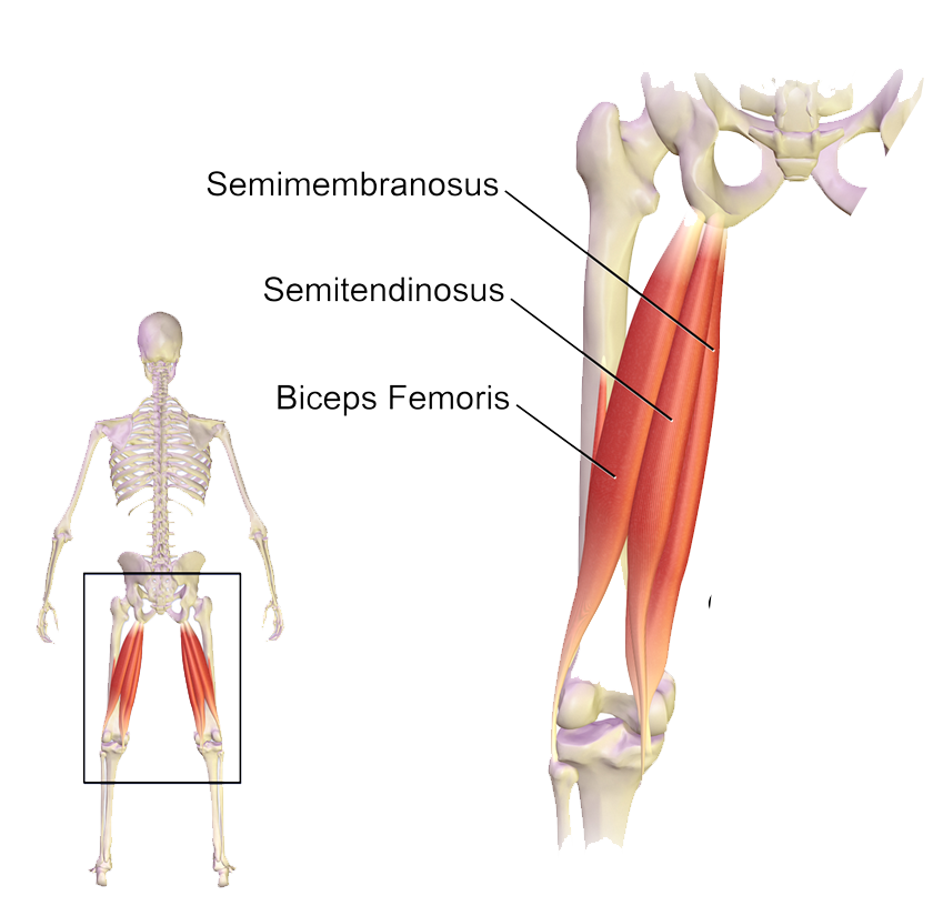
This woman is doing a stretch for the muscles on the back of her legs, the hamstrings. Tibialis anterior, extensor hallucis longus, extensor digitorum longus, fibularis tertius lateral group: Bones are cleverly designed to allow movement at the joints and provide great stability. Skeletal muscles are attached to the bones by the tendons together the skeletal and muscular system allow your body to move. Gastrocnemius muscle anatomy 17 photos of the gastrocnemius muscle anatomy deltoid muscle anatomy, gastrocnemius muscles, gracilis muscle anatomy, plantaris muscle anatomy, quadriceps muscle anatomy, sartorius muscle. Without bones, muscles, and joints, we couldn't stand, walk, run, or even sit. Human muscle system, the muscles of the human body that work the skeletal system, that are under voluntary control, and that are concerned with movement, posture, and balance. Skeletal muscles are attached to the bones by tendons. Skeletal muscles are comprised bundles of muscle fibers. Time to jump right into the biggest and strongest bones in the human body. Editor · aug 13, 2017 ·. Basic muscles and bones of the human body. But how do muscles make your bones move?
Healthy muscular structure and bones. Terms in this set (25). Skeletal muscles are comprised bundles of muscle fibers. When your muscles contract, they pull the bone they're. Tibialis anterior, extensor hallucis longus, extensor digitorum longus, fibularis tertius lateral group:
Muscles Of The Hips And Thighs Human Anatomy And Physiology Lab Bsb 141 from s3-us-west-2.amazonaws.com Learn how to draw the femur, patella, tibia, and fibula in this lesson! The bones of your leg have roughened patches on their surfaces where muscles are attached. Skeletal, smooth and cardiac most skeletal muscles are attached to two bones across a joint, so the muscle serves to move parts. Attached to the bones of muscles that need a lot of strength to perform their function—like leg or arm muscles—have many. Editable vector illustrator cc file (editable live text)editable vector. Fibularis longus, fibularis learn more about the leg and knee anatomy by taking our special quiz, customized to focus on bones, muscles, nerves and vessels of this region! This woman is doing a stretch for the muscles on the back of her legs, the hamstrings. Connecting the pelvic girdle to the lower leg is a bone in the thigh area called the femur, the longest and strongest in the body.
Bones are cleverly designed to allow movement at the joints and provide great stability.
A voluntary muscle usually works across a joint. The sacrum bone is almost always noticeable, no matter what the body type, because it is not covered with muscles or the following life study lower torso and legs in a frontal view, shows the lower torso of a male figure. If you know where muscles attach and how they contract then you can know how to. Gastrocnemius muscle anatomy 17 photos of the gastrocnemius muscle anatomy deltoid muscle anatomy, gastrocnemius muscles, gracilis muscle anatomy, plantaris muscle anatomy, quadriceps muscle anatomy, sartorius muscle. Created and produced by qa international. Muscles can only contract and must work in pairs. The bones of the leg are the femur, tibia, fibula and patella. This woman is doing a stretch for the muscles on the back of her legs, the hamstrings. Skeletal muscles are comprised bundles of muscle fibers. Skeletal muscles are attached to the bones by tendons. Without bones, muscles, and joints, we couldn't stand, walk, run, or even sit. Here we explain the major muscles of the human body. The bones of your leg have roughened patches on their surfaces where muscles are attached.
Bones are cleverly designed to allow movement at the joints and provide great stability leg bones diagram. Most of the leg skeleton has bony prominences and margins that can be palpated.
Share :
Post a Comment
for "Leg Bones And Muscles Diagram / Diagram Illustrating Muscle Groups On Front Of Human Legs Greeting Card By Stocktrekimages Redbubble"


Post a Comment for "Leg Bones And Muscles Diagram / Diagram Illustrating Muscle Groups On Front Of Human Legs Greeting Card By Stocktrekimages Redbubble"